Tuesday, November 27, 2012

REVIEW: 3D Brain App (v1.1)

This free App is available for both the iPhone and the iPad as well as Android Phones.  It was created by Cold Spring Harbor Laboratory (CSH), so my initial expectation was that it would be a good resource.  I highly recommend the  CSH Genes to Cognition website  for students interested in learning about genetic disorders of the brain that influence behavior and cognition.

At the time I downloaded and tested it, the average review it received was 3.5 of 5 stars.

This APP is graphically attractive and the ability to view relatively large anatomical structures in 3D and interior structure through a translucent image of overlying landmarks should be very helpful to those who find traditional 2D drawings meager learning tools.
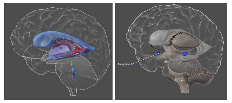

 
     Translucent brain structures help in placing anatomical structures (cerebral ventricles on the left and amygdala on the right) within the brain in relation to overlying landmarks.

The images may be viewed with labels or without; a feature that should be helpful to anyone who is reviewing material for an exam or quiz.  In addition to the images there is an INFO tab that provides a brief but very informative description of the functional aspects of each structure.  Structures can be located either by browsing through a table of contents or be entering a search term memory.  Searches can be done based upon associated functions (e.g., memory, depression) as well as the names of brain structures.

I particularly like that information is provided summarizing research findings regarding the associated functions of the structures and that there are hyperlinks to these primary sources as well as other related internet sites.  In some cases there are case reports that are provided which offer additional evidence for the associated functions of various brain structures (e.g., the patient S.M. and emotional processing within the Amygdala).

The list of structures provided on the iPone App appears to be as comprehensive as the list on the CSH website.

There are more detailed anatomical resources on the web but those can come at a signifcant cost.  This free application should be more than adequate for students in an introductory psychology or neuroscience course.

BOTTOM LINE: Highly Recommended for students in an introductory high school or college psychology or neuroscience course. But be sure to visit the CSH website which is a much more comprehensive resource that will be helpful to students in more advanced courses.

LINK:  The CSH 3D Brain App is available at the Apple App Store and the Android Market.


Wednesday, November 14, 2012

Apps for Psychology & Neuroscience

There has been an explosion of educational Apps for the iPad/iPhone and other similar electronic devises that are being created for a wide variety of academic disciplines.  I thought it might be a useful service to occasionally review some of these here in this Blog.  In addition, if anyone uses an App that I review or finds another that they have used for their psychology or neuroscience courses and found it helpful, please post a comment about the App on this Blog.

While it is strictly speaking not an App, the Khan Academy videos are quite popular.  So to begin, here is my first review of units related to the nervous system, beginning with the basic structure of the neuron.


By this time I often feel that just about every student in my psychology courses has had to at one time learned about the basic structure of the neuron either in middle school or high school.  Nevertheless, instructors religiously review this information and occasionally I see some student examinations/essays that contain inaccuracies. 

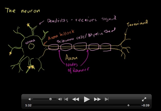
The unit on the neuron in the Khan Academy is essentially accurate, however there are just a few things that I believe could be corrected, missing or could be expanded upon.  

  • While the myelin sheath that insulates some axons is comprised of glial cells called Schwann cells, there are also some myelinated axons in which the sheath is comprised by a second type of glial cell -- oligodendroglia.  Neurons within the central nervous system (brain and spinal cord) are myelinated by oligodendroglia and neurons in the peripheral nervous system are myelinated by Schwann cells.  
  • The Schwan cells and oligodendroglia form myelin in slightly different ways that have important consequences for their function and how susceptible they are to neurodegenerative disease.
  • Khan does not mention that there are non-myelinated axons as well.  These axons are typically extend over very short distances compared with myelinated axons.
  • While were on the topic of the length of axons, Khan mentions that some myelinated axons may attain length of "several feet".  The longest nerve in the human body is the sciatic nerve, a peripheral neuron that extends from  the base of the spine to the big toe of each foot. Injury of this nerve can result in sciatica characterized by  pain, weakness, tingling or numbness in the leg, foot or toes. [BONUS: It is a mixed nerve; one that contains both sensory and motor fibers]. So it would seem logical that the sciatic nerve contains the longest motor axons (maybe 0.5-1 meters depending upon the height a person attains in adulthood).    In his blog Oscillatory Thoughts, Bradley Voytek speculates that sensory sciatic neurons may have even longer axons - ones that might extend from the lower spinal cord at least as high as the brain stem. By the way, using similar logic, the sensory neurons of the sciatic nerve may also have the longest dendrites!  What animal is likely to have the longest axon?  Voytek suggest it would likely be the blue whale.
  • The neuron that is in drawn is the quintessential multipolar motor neuron.  This is generally the image of a neuron that you find in most introductory psychology or neurobiology textbooks.  But   there is a vast array of different types of neurons with different arrangements of their dendrites, soma and axon.  Most intermediate level textbooks also distinguish between bipolar, unipolar, multipolar neurons.


BOTTOM LINE:  This Khan unit is a good review of the basic structural plan of the typical multipolar motor neuron for middle and high school students.  Some information that is likely to be required in introductory and higher level college courses is omitted.

LINK:  Khan Academy Anatomy of a Neuron


Friday, October 19, 2012

Annual Meeting Logo


Back some time ago I challenged readers of this blog to identify what it is that the logo of the SfN meeting represent.


THE ANSWER:  A Tyrosine Kinase Receptor (TRK)


Some members of this diverse family of receptors mediating the response to neurotrophic factors -  molecules that play important roles in determining the development and survival of neurons. The effects of neurotophins are chiefly mediated by TRK-A, TRK-B, and TRK-C receptors.


For example, Neuronal Growth Factor (NGF) is a neurotrophic factor that plays a role in promoting the survival of neurons through the TRK-A receptor.  Interestingly,  NGF can also bind p75 receptors  - a member of the Tumor Necrosis Factor (TNF) family of receptors.

Examine the SfN logo again.  One of the interesting aspects of the TRK family or receptors is that when their ligand (e.g., NGF) is bound to the receptor, they form functional dimers - two paired receptor components.  The function of the activated dimer-receptor complex  is determined by the pair of components that form the dimer.  There can be functional homo-dimers as well as functional hetero-dimers (two similar components or two different components, respectively). An example is p75/TRK-A dimers which are functionally more responsive to NGF than TRK-A homo-dimers (Baker, 2004), .

Another neurotophin, Brain Derived Neurotrophic Factor (BDNF), exerts its influence via the TRK-B receptors.   Chronic stress decreases BDNF production which has been implicated in several neurodegenerative disorders and some psychiatric conditions that are characterized by regionally selective neuronal degeneration such as chronic depression and Post Traumatic Stress Disorder (e.g., Dell'Osso, L. et al, 2009) .
 

For more on BDNF and TRK receptors and depression, you can peruse another blogger's post by following this link, or you can read the review by Nobel et al (2011) - Caution:  the content is comprehensive and advanced).


Cited Sources

Wednesday, October 17, 2012

FUN Poster Session & Social

Monday night  I attended  the annual poster session and social event sponsored by the Faculty for Undergraduate Neuroscience (FUN). Over the past decade the event has grown to be one of the largest of the socials sponsored by the Society for Neuroscience.  There were 165 scientific poster presentations by undergraduates at the social. 

For students, FUN provides competitive travel awards that assist by covering some expenses for undergraduates presenting their research at the conference.  This year there were 16 travel award recipients.  It is also a great opportunity for undergraduates to speak with representatives of graduate programs in neuroscience and for faculty to re-connect with their former students who are now pursuing careers in the neurosciences.  

A number of awards are announced at the FUN social, among these were the Educator of the Year Award, which was awarded to Patsy Dickinson of Bowdoin College (ME), and the Lifetime Achievement Award, presented to Stephen George of Amherst College (MA).

Another important announcement was that The Journal of Undergraduate Neuroscience Education (JUNE) - an open-access, peer-reviewed journal, edited by FUN members - will be listed on PubMed.  The most recent issue of JUNE was just posted on the journal's website: http://www.funjournal.org/  In fact, I have an article on design and assessment of undergraduate neuroscience curricula that is co-authored with colleagues at Ithaca and Oberlin College.

Following the FUN social Nikki Scutella ('10) and I walked a few blocks to enjoy a memorable dinner and get re-acquainted.  Nikki is currently a research assistant in a laboratory of Dr. Gary Fiskum at the University of Maryland Medical College.