Tuesday, December 18, 2012

Primate Midlife Crisis

Increasingly, evolutionary psychologists have join with biologists to explore the relative universality of various phenomenon across the varied branches of the evolutionary tree.  An example of this was recently published in the Proceedings of the Academy of Sciences - USA.  In their study, investigators from several primate research centers across the globe joined forces to ask a simple question - Does it appear that various non-human primates experience a midlife crisis just as many humans do.  The answer they arrived at was  yes they do.

What evidence did they obtain that led them to arrive at this conclusion? First you should know what evidence is there that humans experience a midlife crisis.  When many people think of a midlife crisis they often imagine the middle-aged male whose behaviors may begin to resemble attempts to relive experiences that he had or had wished he had as a younger man.  Similarly, the behaviors of some middle-aged women in the midst of a crisis may also seem to be attempts to recapture their youth. Importantly, not every person may experience this midlife event or if they do, it may not manifest in the same way in every person.  Moreover, what most researchers consider a midlife crisis is much different that what is commonly portrayed in popular media.

What investigators have tended to focus upon as a hallmark of a personal crisis in midlife is a dip in subjective happiness or "well-being".  The data collected are from surveys that include such questions as:  How happy are you with your life?   and How stressful is your life?  An example of the typical pattern of results obtained from individuals of different ages is summarized below from a study by Stone et al. (2010).  Note the dip in overall assessment of subjective well-being (WB) expressed both by men and women during the 4th and 5th decades of life.  [My students might be more concerned about the precipitous decline in WB that appears to occur between the ages of 18 and 25!]


Compare the results above to those obtained for the WB or some of our close primate relatives in the graphs below from the recent paper by Weiss et al. (2012).


There appears to be a clear dip in their assessed WB between the ages of 20 and 40.  But perhaps it would be wise to delve a litter further into the details of the study before we accept that our primate relatives experience a midlife-crisis.

First, your probably thinking it unlikely that the data Weiss et al. obtained was based upon  questionnaires completed by the primates.  Indeed, the assessments of WB were subjective, but they were assessments that were made by humans who were most familiar with the individual animals included in the study.  The raters were zoo keepers, volunteers, researchers, and caretakers who knew the animals for 2 years or more. These humans answered a 4-item survey in which they judged the animals mood (+ vs -), how pleasurable the animal experiences in social interactions, how successfully the animal was at achieving its goals, and how happy the human would be if they were the subject for a week. 

So the data from which the conclusions of the study are drawn are human assessments of the subjective WB of the animal and includes an item that required the human making the assessment to imagine themselves to be the primate they were assessing.  This got me thinking.....

1) How well is any human, even one familiar with primates, accurately able to assess the subjective WB of another human, let alone the WB of a primate?

2) The raters obviously know how old the primates they rated were.  Might  that knowledge play a role in determining their assessments of WB?

Would you consider these significant limitations of the study? Please feel free to share your thoughts.

What I found most interesting about the study was the discussion in which the authors review some of the theories for why there is a "U" shaped age-related pattern to subjective WB in humans.


"The midlife dip cannot be explained by the effects of having young children in the household, and it is similar in males and females, so is not likely connected to menopausal changes or to societal sex roles. A selection explanation, because of the greater longevity of happy people, is likewise unable to account for the midlife dip. One socioeconomic theory is that the U shape reflects hedonic adaptation in which impossible aspirations are first painfully felt around midlife and then slowly and beneficially given up. Another theory is that the curve is linked to financial hardship and thus likely to be less pronounced in those older individuals with higher resources. A third theory is that human aging may bring with it the ability to experience less regret. In short, there is little convergence of explanations about the U-shapes origins." (p. 19949)

In their discussion the authors do suggest that happiness may contribute to longevity and that this may account for the upswing in WB the primate populations they studied.  They also suggest that elevated WB may be adaptive to individuals in their early and later years or that elevated subjective WB may be somehow maladaptive during midlife.

It may also be helpful to consider the individual frame of reference for assessing subjective WB, whether the individual is human or a social primate.  In midlife comparisons may be made both retrospectively and prospectively - against what life had been like when younger and what it could be as compared to others of a similar age group as well as to elders.  Perhaps from this perspective subjective well being is less than desirable.  By contrast as a youth prospects may appear to be quite good and at an advanced age WB may be judged chiefly from what has been accomplished across a lifetime (this seems similar to the socioeconomic theory mentioned above.) In either case subjective WB may be enhanced.

From a neuroscience perspective the authors mention that the age-related "U" shaped pattern of WB may be attributed to maturational changes that occur in various  regions of the brain.  As an example, they site a 2004 study by Urry et al. that compared age correlated age  related difference in WB with activation patterns in the frontal lobes of the brain. They found that enhanced activation of the frontal lobe in the left hemisphere was positively correlated with positive subjective WB (Note: The range of ages of their participants.was between 57 and 60 years).  Left unanswered is if a similar result would also be found among participants in older and younger cohorts.

So at least at my age things appear to be looking up with regard to my subjective WB  ; )

Sources


Tuesday, December 4, 2012

Is the Quality of an Orchestra's Performance Attributable to Mirror Neurons?

Members of a orchestra in the midst of a performance of a symphony by any of your favorite composers must all be attuned to the instrumentation of other performers as well as their own contributions.  For example they must anticipate their own upcoming parts as well as those of the other performers.  The conductor's role in helping keep all the musicians in sync is highly regarded by some, but questioned by others. Couldn't the orchestra perform just as well without a conductor?  Alternatively, should the conductor deserve most of the credited for the distinctive character of what have been the most highly regarded orchestras?   Perhaps both the orchestra members and the conductor deserve equal credit for their most exceptional performances. What, if any information can neuroscientists contribute  that might help answer these questions? 

A report on NPR may be of interest.  In the recent segment, Do Orchestras Really Need a Conductor?, Shankar Vedantam  interviewed Yiannis Aloimonos from the University of Maryland to learn what he and his colleagues found when they conducted experiments to learn what the conductor contributes to the performance of the orchestra. 

In a study, published in the open access journal PlosOne, the investigators were able to map the degree to which the orchestra and conductor were in sync via sensors placed on the ends of the violinist's bows and the conductor's wand.  They asked two different conductors to lead the same orchestra in five different pieces (* see my note at the bottom of this post).  In addition to the data they obtained from the sensors,  they asked experts to judged the subjective quality of the performances.

The statistical analysis of the data obtained from the sensors is complicated, but essentially it allowed the investigators to assess just how well in sync the musicians and conductor were as well as how much "control", "influence" or "drive" the conductor had on the musician's performance.

Here is the briefest of summaries of the outcome:
(Asterisks indicate statistically significant differences between C1 and C2)
  • The two conductors exhibited different degrees of influence over the orchestra (C1 > C2)
  • Under one conductor (C2), the musicians appear to have had a greater influence on each other while playing 3 of 5 pieces.  
  • In 2 of the 5 pieces that the orchestra played, the subjective quality of the performance differed depending upon the conductor; in one instance the price led by conductor 1 prevailed and in the other the performance of the other conductor was judged to be better.
The authors conclude that "... appreciation of (the) music orchestras’ performance was associated to the concurrent increase of conductor-to-musician influence and a reduction of musician-to-musician information flow. " (p. e35757).

In other words, the conductor does have an influence on the aesthetic quality of the performance as long as her/his contributions  "drive" the performance of the orchestra and that the relative influence of other musicians on each other's performance is secondary to that of the conductor's.  

Examine the graphs above and below carefully.  Do they appear to support the authors' conclusions? 


(Asterisks indicate statistically significant differences between C1 and C2)

Statistically the there appears to be a significant difference in the drive exerted on the orchestra by the two conductors during performance of pieces 3 and 5 (C1 > C2) and likewise, musician-to-musician influence was greater during performance of pieces 1, 2, and 3 under the direction of C2.  Differences in subjective aesthetic perception of the performances were significant for piece 3 and 5.  In both instances, C1 was able to exert a greater influence on the musicians. However in one instance the subjective ratings of the performance under direction by C1 were better (Piece 3) and in the other instance the performance under the direction of C1 were judged to be poorer. Obviously there seems to be some complex interaction of factors that is occurring.  


What the authors of the paper argue is that in the instance of Piece 3, the relatively greater degree of direction achieved by C1 in combination with diminished musician-to-musician influences resulted in a more aesthetic performance than what was achieve by C2.

So far so good.  But what explains the poorer performance of Piece 5 under the direction of C1, despite achieving relatively greater drive than C2?  The authors argue that despite exerting greater drive, C1 did not decrease the musician-to-musician influences any more than C2.

Altogether, this pattern of results supports the conclusions of the researchers.

But might there be more at play here?  Is it possible that the piece itself may also play a role in whether the drive achieved by the conductor  in combination with relatively less musician-to-musician is ultimately what results in an aesthetically pleasing performance? The authors admit as much, but future research will be needed to assess the influence of the musical score itself.

Why might any of this interest a neuroscientist, even those neuroscientists who do not regularly attend orchestral performances?

What might be going on in the brain of the musicians and the conductor that interests a neuroscientist?   Although it is highly speculative, the authors of the paper suggest that  the interaction between conductor and the orchestra.

Mirror neurons are neurons that are activated when an individual engages in a specific action or observes another individual engage in that same action.   Mirror neurons were first identified by researchers recording from individual neurons within the brain's of monkeys (the goal of this work was to determine if with training, the monkeys could remotely control the movement of a prosthetic arm and hand using only the input from these neurons). However subsequent neuro-imaging studies with humans have identifies networks with some brain regions that appear to function similarly to mirror neurons.

Presumably the conductor's influence on the members of the orchestra may be mediated by networks comprised of mirror neurons. It also seems likely that  the musician-to-musician interactions might be mediated by such networks. What may matter aesthetically to the audience is that the influence of the conductor dominates. 

What is true for the orchestra may also be true for the chorus! Let me know what you think!

In a study published in the journal Science in 2002 the investigators demonstrated that some mirror neurons of monkeys responded most strongly to the the sight of actions paired with the sounds that the actions caused (i.e., paper being ripped, a stick being dropped).  Might this be comparable to the gestures of the conductor and the resulting changes in the musical passage that they are intended to signal?  I was also reminded of a study published in the journal Nature a few years ago in which neuroscientists studying the neural circuits that control the songs of some birds report evidence of mirror-neuron-like activation of the song system when the birds are exposed to the characteristic songs of their own species, but not distinctive songs of a another species of songbird.  These studies were supposedly the first to find evidence of mirror neuron networks in a non-primate species.

Mirror neurons have received a great deal of attention over the past decade. They have been implicated as serving a role in observational learning and in empathic perception.  One theory that has garnered a great deal of interest is that autistic individuals may have deficits in the mirror neuron circuitry.

NOTE:  In actuality the study did not employ an entire orchestra, only 8 violinists.   It is also not clear specifically what musical pieces were used, although the authors mention Mozart Symphony No. 40

Sources
  • D'Ausilio A, Badino L, Li Y, Tokay S, Craighero L, et al. (2012) Leadership in orchestra emerges from the causal relationships of movement kinematics. PLoS ONE 7(5): e35757. doi:10.1371/journal.pone.0035757
  • Iacoboni M, Molnar-Szakacs I, Gallese V, Buccino G, Mazziotta JC, et al. (2005) Grasping the intentions of others with one’s own mirror neuron system. PLoS Biol 3(3): e79.
  • Kohler, E., et al. (2002).  Hearing sounds, understanding actions: Action representation in mirror neurons. Science, 297, 846-848.
  • Miller, G. (2008). Mirror neurons may help songbirds stay in tune.  Science, 319, 269.
  • Ramachandran, V.S. & Oberman, L.M. (November, 2006).  Broken mirrors. A theory of autisms.  Scientific American, 295 (6), 63-69.
  • Rizzolantti, G., Fogassi, L., & Gallese, V. (November, 2006).  Mirrors in the minds.  Scientific American, 295 (6), 54-61.


Tuesday, November 27, 2012

REVIEW: 3D Brain App (v1.1)

This free App is available for both the iPhone and the iPad as well as Android Phones.  It was created by Cold Spring Harbor Laboratory (CSH), so my initial expectation was that it would be a good resource.  I highly recommend the  CSH Genes to Cognition website  for students interested in learning about genetic disorders of the brain that influence behavior and cognition.

At the time I downloaded and tested it, the average review it received was 3.5 of 5 stars.

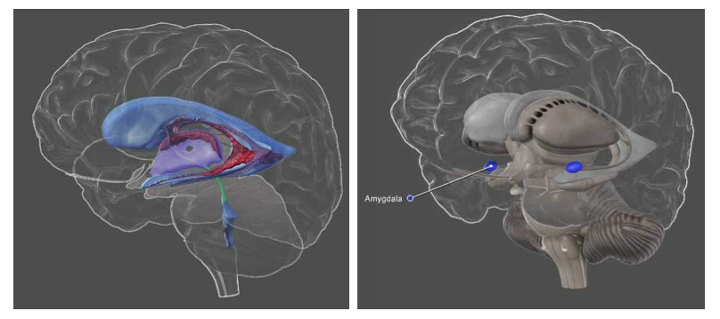
This APP is graphically attractive and the ability to view relatively large anatomical structures in 3D and interior structure through a translucent image of overlying landmarks should be very helpful to those who find traditional 2D drawings meager learning tools.


     Translucent brain structures help in placing anatomical structures (cerebral ventricles on the left and amygdala on the right) within the brain in relation to overlying landmarks.

The images may be viewed with labels or without; a feature that should be helpful to anyone who is reviewing material for an exam or quiz.  In addition to the images there is an INFO tab that provides a brief but very informative description of the functional aspects of each structure.  Structures can be located either by browsing through a table of contents or be entering a search term memory.  Searches can be done based upon associated functions (e.g., memory, depression) as well as the names of brain structures.

I particularly like that information is provided summarizing research findings regarding the associated functions of the structures and that there are hyperlinks to these primary sources as well as other related internet sites.  In some cases there are case reports that are provided which offer additional evidence for the associated functions of various brain structures (e.g., the patient S.M. and emotional processing within the Amygdala).

The list of structures provided on the iPone App appears to be as comprehensive as the list on the CSH website.

There are more detailed anatomical resources on the web but those can come at a signifcant cost.  This free application should be more than adequate for students in an introductory psychology or neuroscience course.

BOTTOM LINE: Highly Recommended for students in an introductory high school or college psychology or neuroscience course. But be sure to visit the CSH website which is a much more comprehensive resource that will be helpful to students in more advanced courses.

LINK:  The CSH 3D Brain App is available at the Apple App Store and the Android Market.


Wednesday, November 14, 2012

Apps for Psychology & Neuroscience

There has been an explosion of educational Apps for the iPad/iPhone and other similar electronic devises that are being created for a wide variety of academic disciplines.  I thought it might be a useful service to occasionally review some of these here in this Blog.  In addition, if anyone uses an App that I review or finds another that they have used for their psychology or neuroscience courses and found it helpful, please post a comment about the App on this Blog.

While it is strictly speaking not an App, the Khan Academy videos are quite popular.  So to begin, here is my first review of units related to the nervous system, beginning with the basic structure of the neuron.


By this time I often feel that just about every student in my psychology courses has had to at one time learned about the basic structure of the neuron either in middle school or high school.  Nevertheless, instructors religiously review this information and occasionally I see some student examinations/essays that contain inaccuracies. 

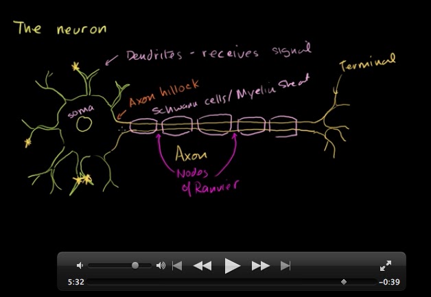
The unit on the neuron in the Khan Academy is essentially accurate, however there are just a few things that I believe could be corrected, missing or could be expanded upon.  

  • While the myelin sheath that insulates some axons is comprised of glial cells called Schwann cells, there are also some myelinated axons in which the sheath is comprised by a second type of glial cell -- oligodendroglia.  Neurons within the central nervous system (brain and spinal cord) are myelinated by oligodendroglia and neurons in the peripheral nervous system are myelinated by Schwann cells.  
  • The Schwan cells and oligodendroglia form myelin in slightly different ways that have important consequences for their function and how susceptible they are to neurodegenerative disease.
  • Khan does not mention that there are non-myelinated axons as well.  These axons are typically extend over very short distances compared with myelinated axons.
  • While were on the topic of the length of axons, Khan mentions that some myelinated axons may attain length of "several feet".  The longest nerve in the human body is the sciatic nerve, a peripheral neuron that extends from  the base of the spine to the big toe of each foot. Injury of this nerve can result in sciatica characterized by  pain, weakness, tingling or numbness in the leg, foot or toes. [BONUS: It is a mixed nerve; one that contains both sensory and motor fibers]. So it would seem logical that the sciatic nerve contains the longest motor axons (maybe 0.5-1 meters depending upon the height a person attains in adulthood).    In his blog Oscillatory Thoughts, Bradley Voytek speculates that sensory sciatic neurons may have even longer axons - ones that might extend from the lower spinal cord at least as high as the brain stem. By the way, using similar logic, the sensory neurons of the sciatic nerve may also have the longest dendrites!  What animal is likely to have the longest axon?  Voytek suggest it would likely be the blue whale.
  • The neuron that is in drawn is the quintessential multipolar motor neuron.  This is generally the image of a neuron that you find in most introductory psychology or neurobiology textbooks.  But   there is a vast array of different types of neurons with different arrangements of their dendrites, soma and axon.  Most intermediate level textbooks also distinguish between bipolar, unipolar, multipolar neurons.


BOTTOM LINE:  This Khan unit is a good review of the basic structural plan of the typical multipolar motor neuron for middle and high school students.  Some information that is likely to be required in introductory and higher level college courses is omitted.

LINK:  Khan Academy Anatomy of a Neuron


Friday, October 19, 2012

Annual Meeting Logo


Back some time ago I challenged readers of this blog to identify what it is that the logo of the SfN meeting represent.


THE ANSWER:  A Tyrosine Kinase Receptor (TRK)


Some members of this diverse family of receptors mediating the response to neurotrophic factors -  molecules that play important roles in determining the development and survival of neurons. The effects of neurotophins are chiefly mediated by TRK-A, TRK-B, and TRK-C receptors.


For example, Neuronal Growth Factor (NGF) is a neurotrophic factor that plays a role in promoting the survival of neurons through the TRK-A receptor.  Interestingly,  NGF can also bind p75 receptors  - a member of the Tumor Necrosis Factor (TNF) family of receptors.

Examine the SfN logo again.  One of the interesting aspects of the TRK family or receptors is that when their ligand (e.g., NGF) is bound to the receptor, they form functional dimers - two paired receptor components.  The function of the activated dimer-receptor complex  is determined by the pair of components that form the dimer.  There can be functional homo-dimers as well as functional hetero-dimers (two similar components or two different components, respectively). An example is p75/TRK-A dimers which are functionally more responsive to NGF than TRK-A homo-dimers (Baker, 2004), .

Another neurotophin, Brain Derived Neurotrophic Factor (BDNF), exerts its influence via the TRK-B receptors.   Chronic stress decreases BDNF production which has been implicated in several neurodegenerative disorders and some psychiatric conditions that are characterized by regionally selective neuronal degeneration such as chronic depression and Post Traumatic Stress Disorder (e.g., Dell'Osso, L. et al, 2009) .
 

For more on BDNF and TRK receptors and depression, you can peruse another blogger's post by following this link, or you can read the review by Nobel et al (2011) - Caution:  the content is comprehensive and advanced).


Cited Sources

Wednesday, October 17, 2012

FUN Poster Session & Social

Monday night  I attended  the annual poster session and social event sponsored by the Faculty for Undergraduate Neuroscience (FUN). Over the past decade the event has grown to be one of the largest of the socials sponsored by the Society for Neuroscience.  There were 165 scientific poster presentations by undergraduates at the social. 

For students, FUN provides competitive travel awards that assist by covering some expenses for undergraduates presenting their research at the conference.  This year there were 16 travel award recipients.  It is also a great opportunity for undergraduates to speak with representatives of graduate programs in neuroscience and for faculty to re-connect with their former students who are now pursuing careers in the neurosciences.  

A number of awards are announced at the FUN social, among these were the Educator of the Year Award, which was awarded to Patsy Dickinson of Bowdoin College (ME), and the Lifetime Achievement Award, presented to Stephen George of Amherst College (MA).

Another important announcement was that The Journal of Undergraduate Neuroscience Education (JUNE) - an open-access, peer-reviewed journal, edited by FUN members - will be listed on PubMed.  The most recent issue of JUNE was just posted on the journal's website: http://www.funjournal.org/  In fact, I have an article on design and assessment of undergraduate neuroscience curricula that is co-authored with colleagues at Ithaca and Oberlin College.

Following the FUN social Nikki Scutella ('10) and I walked a few blocks to enjoy a memorable dinner and get re-acquainted.  Nikki is currently a research assistant in a laboratory of Dr. Gary Fiskum at the University of Maryland Medical College. 

What does the SFN Conference Logo Represent?

Prior to the SfN conference I challenged readers to try and identify what it is that is represented in the SfN Conference Logo.   Tomorrow I'll post the answer.So there is still time to submit your answer as a comment to this post.

Here are some hints:
  • It is found in the cell membrane of some neurons.
  • It has a ligand binding site - a site where chemical messengers (e.g., neurotransmitters) can bind.
  • It is NOT an ionotropic receptor complex

Monday, October 15, 2012

SOMAS Luncheon @ SfN

It was a great privilege to attend the 2012 SOMAS Luncheon at the annual SfN meeting yesterday.  This is a great program that has supported undergraduate neuroscience instructors in the early stages of their careers and provided funding in support of the collaborative student-faculty research.

The 2012 SOMAS award winners and their students are:

  • Carlita Favero, Ph.D.  Ursinus College
  • Elizabeth Glater, Ph.D.  Harvey Mudd College
  • Josef G. Trapani, Ph.D.  Amherst College
  • Charles Weaver, Ph.D.  Saginaw Valley State University
  • Sarah Webster, Ph.D.  College of the Holy Cross

You can learn more about their projects at the SOMAS website: http://www.somasprogram.org/

The Brain on Idle

Sophisticated and highly technical functional brain imaging techniques such as fMRI and PET have revolutionized the cognitive and behavioral neurosciences.  These techniques offer a valuable non-invasive means for visualizing, localizing, and quantifying patterns of brain activity.  But a recent essay in the journal Nature focuses on what might be the important functions of the baseline brain activity that occurs when subjects in these scanners are instructed to "clear your mind".  In such instances brain activity diminishes by just a 1-5%. Some investigators believe that there is much to be learned from examining the resulting patterns in activity in the idling brain (mind). Here are some suggestions that are presented by the essays author,  Kerri Smith:
  • idling brain activity may keep essential neural pathways interconnected.
  • the activity may be necessary to prime the brain for action
  • it might be performing essential "off-line" functions related to such processes as memory consolidation.
  • it may even be that this activity may be "meaningless" - e.g., fMRI actually measures patterns of regional blood flow, not neural activity itself.
However, according to Smith, there is general agreement among most investigators that these are mere guesses (hypotheses).  What is more, it is not clear how to devise experiments to test these alternative hypotheses.

Here are some other facts that are shared in the essay:
  • patterns of intrinsic brain activity differ in distinctive ways between healthy controls and individuals with various brain disorders such as Alzheimer Disease and Autism.
  • distinctive patterns of intrinsic brain activity at any instant might predispose individuals to respond to ambiguous situations or stimuli. 
To illustrate the latter point, Smith summarizes the result of a study (Hesselmann et al., 2008) that is somewhat similar to another I learned about in a recent presentation at the College by Bill Klemm of Texas A&M.  Hesselmann et al.  compared the intrinsic patterns of brain activity in subjects just before they were presented with some classic ambiguous visual stimuli.  When presented with such stimuli subjects might perceive either the image of a face or some other image such as a vase or a reclining woman. According to Klemm what a particular individual is more likely to see initially is relatively constant from one trial to the next – the perception that an individual experiences first tend to be  reliably consistent.  Another way to say this is that is seems that their brain is prepared (possibly biased) toward perceptions of a particular nature.  And in the study by Hesselmann et al. the intrinsic pattern of activity observed in those participants who initially perceive a face was characterized by enhanced activity with a region that is known to play a role in facial perception - the fusiform face area (FFA).  The intrinsic activity of their brian just before the stimulus presentation apparently influenced their perceptions.

Are you prepared to step on your accelerator?  What hypotheses might you have for the function of intrinsic brain activity?  Do you anticipate that such intrinsic activity should resemble activity during mediation or sleep? Please share your hypotheses or ideas for how experiments might be performed to test some hypothesis presented above.

Sources Cited
You can listen to a podcast related to this topics here:

Saturday, October 13, 2012

Chuck Close - My Life as a Rolling Neurological Clinic

So what did Close have to share in his public presentation?  

Due to health issues that left Close unable to travel, he appeared via a satellite link from his studio in NYC.   For the first half-hour he shared with us the events and circumstances in his life that shaped his art.  Among these were, poverty his family experienced, the early death of his father, the influence of his grandmother who herself was housebound by agoraphobia, his great difficulty recognizing faces, his learning disabilities, the mid-career collapse of a spinal artery that left him unable to move below his chest  and how it was that art had ultimately "saved his life".   How had art done this?  Because despite what appeared to be limitations, art allowed him to demonstrate that he still had something he could contribute.  There has been something about the accomplishments that Close has attained that has transcended his physical abilities. 

To me what this may illustrate is just how much will remain to be explained by neuroscience even if we ultimately reach a complete understanding of the biological workings of the brain.  While I am usually more comfortable in my own research working from the assumptions of a biological reductionist, the alternative explanations remain both a challenge and an inspiration for pursuing research in the behavioral and neural sciences.  

In my view, there remains no greater challenge in science than to pursue an understanding of brain function.   Yet even should we obtain such an understanding, what might ultimately remain unfathomable will surely be inspiration for the efforts of future neuroscientists.  To be a scientists is inevitably to recognize that each answer that is achieved results in an innumerable number of questions that you've likely to have never contemplated. As Close shared, the process of constructing each of his portraits was often more rewarding than the product.  Another way of expressing this is that the journey is somehow more enlightening than reaching your destination. In my experience, this is almost always the case.

To enhance his journey, at each new phase of Close's career he challenge himself to embark upon a new approach to portraiture - remove color, add it back, add elements ("incremental units") within either a horizontal, diagonal or radial grid.  In each instance, the challenge that Close set for himself seemed to me to be much like the challenge before each chemists, physicist or neuroscientist.  That challenge is to understand how such incremental units (e.g., each, element, subatomic particle or neuron), which in isolation may seem rather simple, can collectively contribute to something much greater.  

There is also something inspiring to me, and I hope to my students, about how the collective contributions of a single neuroscientist may ultimately result in achieving a much more comprehensive understanding of the brain and mind than would otherwise be attainable.  Today the total number of registrants attending the SfN conference reached nearly 28,000.  By the end of the conference that number will probably exceed 30,000.  That is very encouraging.

Lastly, there are a few things Close shared that piqued my curiosity.  For example, he described his face-blindness  as a difficulty recognizing three-dementional faces.  It was much easier for him to work from two-dimensional photographs of his subjects than the subjects themselves.  One of the great quandaries in neuroscience is the Binding Problem -- i.e., how is it that features (elements) that are represented in disparate regions or neural circuits are somehow combined to create a perceptual unity, e.g., a unique facial percept.

I wonder if there have been any studies that might indicate that face-blindness might arise in some instances from a deficit in a circuit that is responsible for binding "flat" incremental facial features into a three-dimensional facial percept.  Anyone out there know if their is any evidence of this in the neuroscience literature? Notably, most studies of prosopagnosia employ two-deimensional "flat" facial stimuli.  Might this mean that the incidence of face-blindness is even more common than is estimated?

Katrina's Aftermath

For several years the SfN Meeting was held between venues in DC, Florida, St Louis, Los Angeles/San Diego, and New Orleans.   In 197, 2000 and again in 2003 the meeting was to be held in New Orleans.  Katrina changed that. The views you see here describe the path of the hurricane, a view of the populace that sought refuge at the Convention Center and  view from the air of the flooding around the Super-dome which is within a mile of the convention center. 

The 2006 and 2009 meetings were scheduled to return to NO, but the 2005 hurricane caused the society to cancel those plans.The 2012 meeting is the first time that the meeting returns to New Orleans.  It seemed ironic that just a few month ago, NO was under threat of another disaster from hurricane Isaac.

My first impressions upon returning....

Keep in mind that my impressions thus far are limited only to the things I've been able to observe from my arrival yesterday afternoon until this morning.   From what I've seen and done thus far, things seem much the same.  There is a great deal of construction/re-construction that appears to still be going on.  The people I've met so far seem extremely friendly and are very glad that there are visitors form out of town.  The local news last night featured a story on people who were still displaced from their homes by Isaac - an hurricane season is not over.  But for this week, the forecast is for sunny days and mild nights.  I'll add more to this post throughout my visit.  

Tuesday, October 9, 2012

Mysteries of the Brain


In conjunction with the SfN meeting, the journal Science has published a special issue with featured articles on various “Mysteries of the Brain” and on Chronic Depression.  Here is an opportunity to determine the content of one of my posts on this blog.  Please let me know which of the following articles you would like me to feature in the blog. The majority of request I receive via comments prior to Saturday, October 13 will determine what I’ll post in the blog.   Here are the choices: 
  • Why is mental illness so hard to treat?
  • Why are our brains so big?
  • Why are you and your brain unique?
  • Can we make our brains more plastic?
    •